
TETRA Launches New Interactive App for Metamaterial-Informed Tissue Characterization
3 September 2026
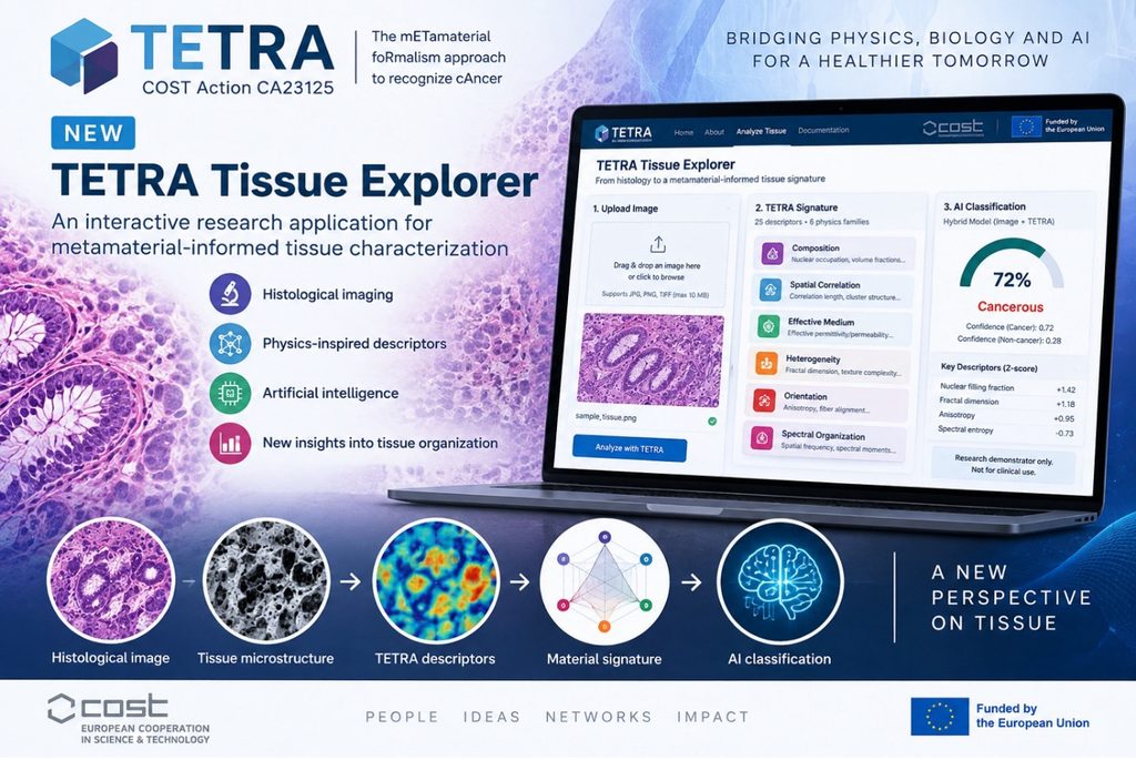
COST Action TETRA (CA23125) is pleased to announce the launch of the TETRA Tissue Explorer, a new interactive research application that demonstrates how concepts from metamaterial physics can be combined with digital histology and artificial intelligence for tissue characterization.
Developed within the framework of TETRA – The mETamaterial foRmalism approach to recognize cAncer – the application translates histological tissue images into quantitative, physics-inspired representations. Rather than treating tissue exclusively as a digital image, the TETRA approach considers its microscopic architecture as a complex structured material.
From a histological image to a TETRA material signature
Users can upload an H&E histological image to the application. The TETRA Tissue Explorer then extracts 25 quantitative descriptors, organized into six physics-inspired families. These characterize aspects of tissue architecture including nuclear occupation, heterogeneity, structural complexity, spatial correlations, anisotropy, spectral organization and effective-medium surrogates.
The result is a distinctive TETRA material signature that provides a new way of representing tissue microstructure.
The underlying concept can be summarized as:
Histology → Microstructure → TETRA descriptors → Effective representation → Physics-informed AI
Bringing physics and artificial intelligence together
A major objective of the demonstrator is to explore whether physics-informed descriptors can provide information complementary to conventional image-based artificial intelligence.
The TETRA framework therefore enables a hybrid approach in which deep-learning features extracted directly from histological images can be combined with the quantitative TETRA material representation. This creates a platform for investigating whether changes associated with cancer can be characterized not only visually, but also through changes in the multiscale physical organization of tissue.
Publicly available colorectal histopathology datasets are being used for model development and independent validation, while future work will extend the approach through TETRA collaborations and appropriately labelled datasets.
Turning the TETRA concept into an interactive demonstrator
The TETRA Tissue Explorer represents an important step in translating the scientific concept behind the Action into a tangible digital tool.
It brings together expertise spanning metamaterials, electromagnetics, biophotonics, digital pathology, image analysis and artificial intelligence, demonstrating the interdisciplinary character of the TETRA network.
The application will also provide a platform for further collaborative research, comparison of conventional and physics-informed AI approaches, and future validation using datasets contributed by TETRA partners.
A research demonstrator
The TETRA Tissue Explorer is currently a research prototype and is not a medical device. Its material parameters are morphology-derived computational descriptors rather than direct measurements of tissue optical or electromagnetic properties, and its outputs must not be used for clinical diagnosis or patient management.
Nevertheless, the demonstrator illustrates the central scientific vision of TETRA: to investigate whether the language and formalism of metamaterials can provide new ways to describe, quantify and ultimately recognize disease-associated changes in biological tissue.
TETRA Tissue Explorer — see tissue through the lens of metamaterial physics.